See 3-D models of animal anatomy from openVertebrate’s public collection
More than 13,000 museum specimens were CT scanned as part of a six-year-long project
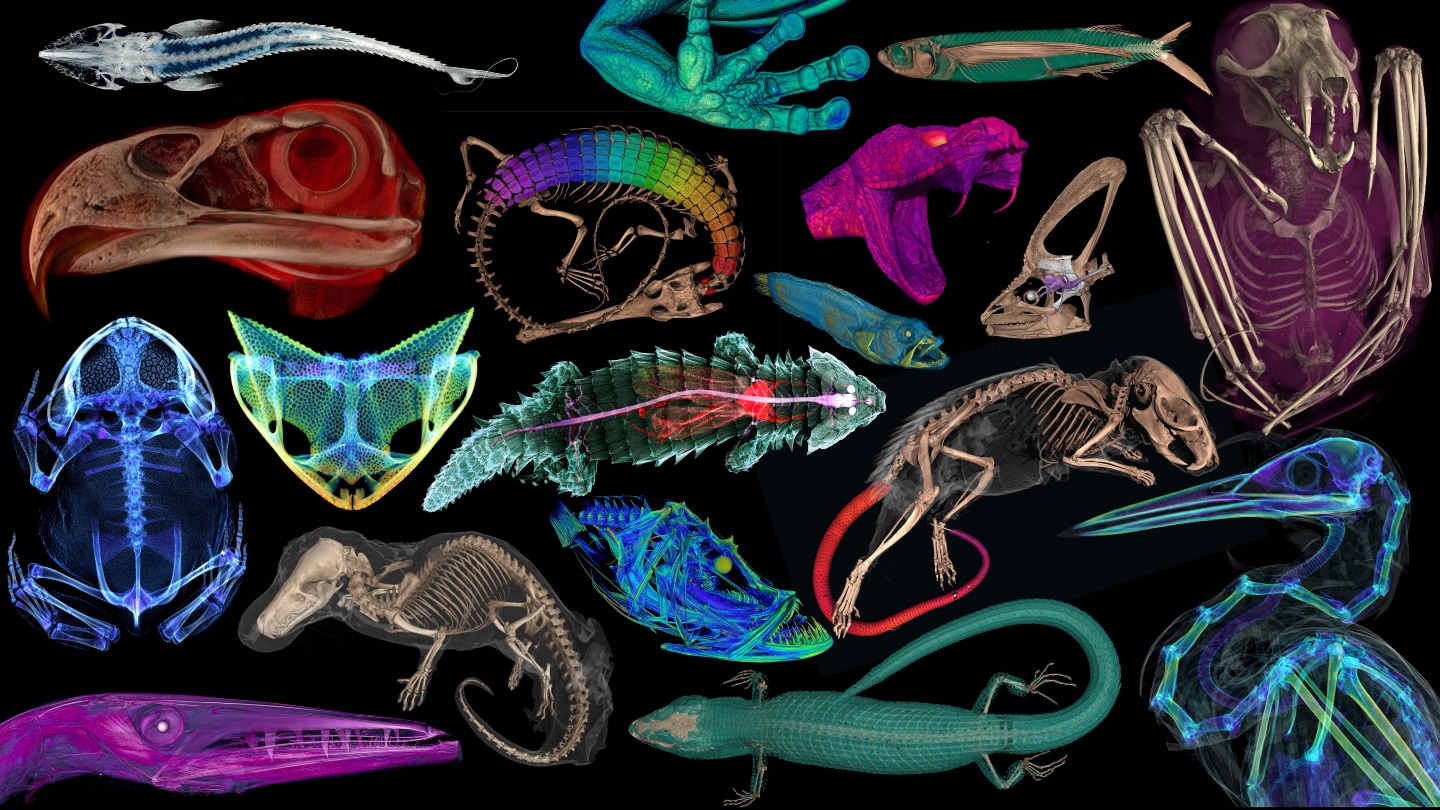
With the completion of a yearslong project called openVertebrate, the insides of more than 13,000 museum specimens are seeing the light of day. Digital reconstructions of CT scans (some shown) show the anatomy of fluid-preserved vertebrates, as well as last meals, yet-to-be-born offspring and more.
openVertebrate